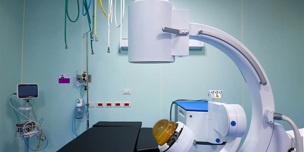
Cardiovascular disease (CVD) is the leading cause of death worldwide, accounting for approximately 31% of all deaths globally. Early detection of CVD is crucial in preventing its progression and associated complications. Non-invasive diagnostic techniques are important tools for detecting early-stage CVD. In this blog post, we will discuss advancements in non-invasive diagnostic techniques for detecting early-stage CVD.
Non-invasive diagnostic techniques refer to methods that do not involve the insertion of instruments or devices into the body. These techniques are safe, relatively low-cost, and can be easily repeated over time. Non-invasive techniques are particularly useful in detecting early-stage CVD, as they can identify subtle changes in the structure and function of the heart and blood vessels before symptoms appear.
Echocardiography uses high-frequency sound waves to produce images of the heart. This technique can be used to visualize the heart's structure and function and is particularly useful in detecting abnormalities such as valve disorders, heart failure, and congenital heart defects. Echocardiography is non-invasive, safe, and has no known risks or side effects
Electrocardiography measures the electrical activity of the heart and can be used to detect abnormalities such as arrhythmias, ischemia, and heart attacks. ECG is non-invasive, painless, and does not involve any radiation exposure. ECG is often used as a routine screening tool in patients with risk factors for CVD.
Magnetic Resonance Imaging uses a strong magnetic field and radio waves to produce detailed images of the heart and blood vessels. MRI is particularly useful in detecting abnormalities such as aneurysms, dissections, and myocardial infarctions. MRI is non-invasive, does not involve radiation exposure, and is considered safe for most patients.
Computed Tomography uses X-rays to produce detailed images of the heart and blood vessels. CT scans can be used to detect abnormalities such as coronary artery disease, pulmonary embolism, and aortic aneurysms. CT scans are non-invasive, relatively quick, and do not involve any radiation exposure.
Stress testing involves exercising the heart to its maximum capacity while monitoring its function using ECG or echocardiography. Stress testing can be used to detect abnormalities such as ischemia and arrhythmias. Stress testing is non-invasive, safe, and has no known risks or side effects.
Advancements in non-invasive diagnostic techniques have led to the development of several new technologies that can detect early-stage CVD with even greater accuracy and precision. Some of these technologies include:
Cardiac Magnetic Resonance Imaging uses advanced MRI techniques to produce highly detailed images of the heart and blood vessels. CMR is particularly useful in detecting abnormalities such as myocardial fibrosis and cardiomyopathy. CMR is non-invasive, safe, and has no known risks or side effects
Optical Coherence Tomography uses light waves to produce high-resolution images of the heart's blood vessels. OCT is particularly useful in detecting abnormalities such as plaque buildup and thrombosis. OCT is non-invasive, safe, and has no known risks or side effects
Positron Emission Tomography uses a radioactive tracer to produce images of the heart and blood vessels. PET is particularly useful in detecting abnormalities such as inflammation and atherosclerosis. PET is non-invasive, safe, and has no known risks or side effects, although the use of a radioactive tracer may be contraindicated in some patients with certain medical conditions.
Get premium cardiac diagnostics services in delhi ncr at affordable price just give us call on 9999657744 or mail us on delhicardio@gmail.com